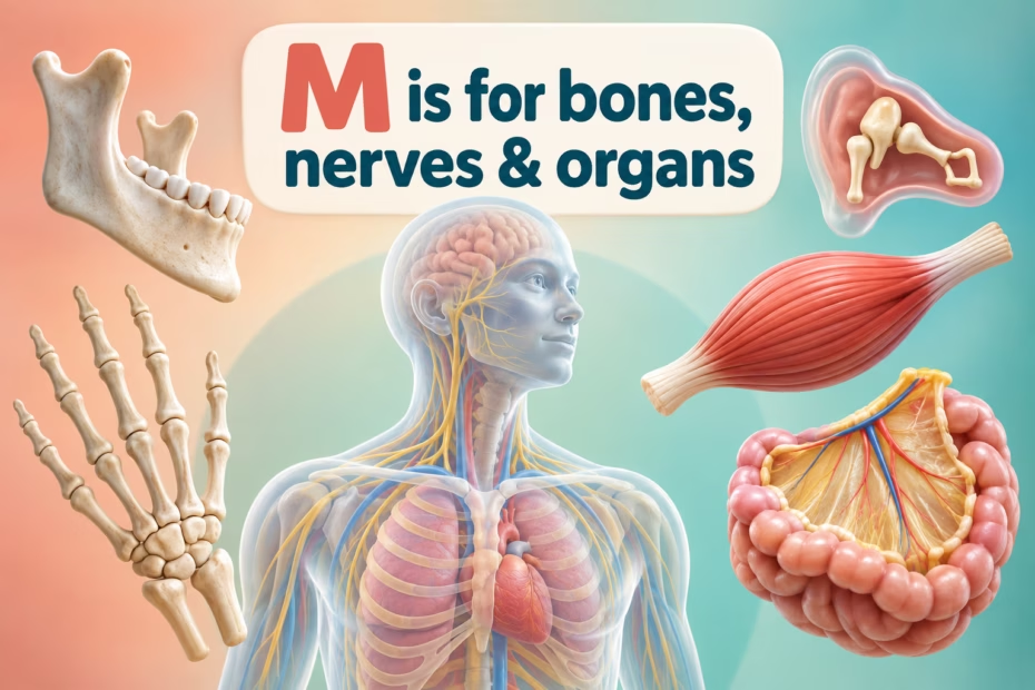
Human body parts that start with M include bones such as the mandible, nerves such as the median nerve, organs such as the myocardium, and regions such as the mediastinum. This alphabetized list mixes structures of different sizes and types, so the entries are vocabulary examples rather than 34 separate organs.
What is included in the M anatomy list?
The directory below includes 34 terms, but they do not all describe the same kind of structure. Some are bones or joints; others are nerves, muscles, body regions, membranes, or larger systems. For instance, the metacarpals are five bones in the palm, while the muscular system names a broad network of skeletal, smooth, and cardiac muscle.
| Type | M examples | How to distinguish them |
|---|---|---|
| Bones and joints | Mandible, maxilla, malleus, metacarpals, metatarsals | Individual bones or groups of bones; the metacarpals are in the palm and metatarsals are in the midfoot. |
| Nerves | Mandibular, maxillary, median, musculocutaneous nerves | Carry sensory information, motor commands, or both; nerve roles are specific rather than interchangeable. |
| Muscles and muscle tissue | Masseter, medial rectus, mylohyoid, myocardium | Muscles contract. Myocardium is the muscular middle layer of the heart wall. |
| Membranes, spaces, and regions | Meninges, mediastinum, middle cranial fossa, maxillary sinus | These name protective layers, compartments, skull-base regions, or air-filled spaces—not separate organs. |
| Organs and systems | Mammary glands, mouth, muscular system | Some entries are organs or glandular structures; a system groups many related tissues and organs. |
How do the malleus and other middle-ear bones help hearing?
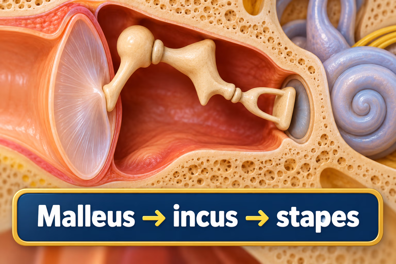
The malleus is one of the three auditory ossicles in each middle ear. Its handle attaches to the eardrum. When the eardrum vibrates, the malleus moves the incus, which moves the stapes; the stapes passes the vibration to the oval window of the inner ear. This sequence is why the malleus should not be described as carrying vibration directly to the inner ear.
Learn more in the Cleveland Clinic overview of the ossicles.
What do the mandibular and maxillary nerves do?
The mandibular nerve (V3) and maxillary nerve (V2) are two branches of the trigeminal nerve. Both carry sensation from areas of the face. V3 is the only trigeminal division with motor fibers: it supplies the muscles of mastication and a few additional muscles. V2 is sensory; it does not control facial expressions or supply taste. Facial-expression muscles are primarily controlled by the facial nerve (cranial nerve VII).
The mandibular nerve’s sensory branches serve areas including the lower teeth, lower lip, chin, and general sensation from the front part of the tongue. Taste fibers from the facial nerve travel alongside the lingual nerve for part of their route, which is different from saying that V3 itself provides taste. The NCBI Bookshelf trigeminal anatomy overview describes the branches and their separate functions.
What does the median nerve supply?
The median nerve travels through the forearm and wrist into the hand. It supplies selected forearm and hand muscles and sensation to much of the palm side of the thumb, index finger, middle finger, and thumb-side half of the ring finger. It does not supply every finger or the entire forearm. At the wrist it passes through the carpal tunnel, where compression can cause carpal tunnel syndrome. See NCBI Bookshelf’s median-nerve anatomy.
What are the mesentery and mediastinum?
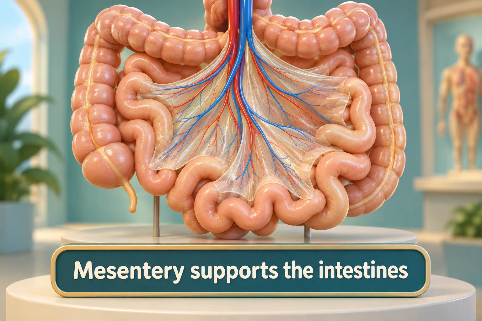
The mesentery is a fold of peritoneum that attaches portions of the intestine to the abdominal wall and carries blood vessels, nerves, and lymphatic vessels. Calling it a “newly discovered organ” is misleading: its anatomy has long been described, while modern discussion has revisited how best to classify and name its continuous structure.
The mediastinum is the central compartment in the chest between the right and left pleural cavities. It contains structures such as the heart, great vessels, trachea, esophagus, and thymus. It is not a sturdy wall that separates the lungs; each lung lies in its own pleural cavity. The NCBI thorax overview describes these compartments.
Where is the medulla oblongata?
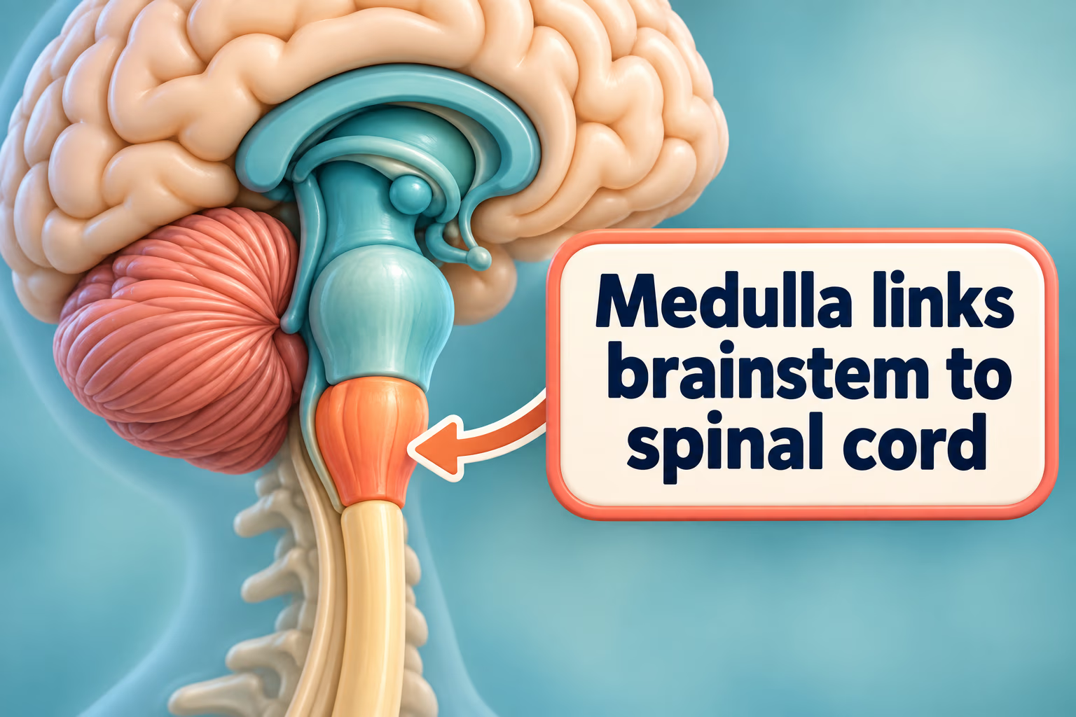
The medulla oblongata is the lowest portion of the brainstem and is continuous with the spinal cord. It contains ascending and descending pathways and centers involved in automatic functions such as breathing and cardiovascular regulation. “Bridge” can be a handy visual shorthand for its location, but the medulla is active nervous tissue, not simply a passive connector.
For an overview, see NCBI Bookshelf’s medulla oblongata reference.
How should other M anatomy facts be read?
- Meninges: dura mater, arachnoid mater, and pia mater surround the brain and spinal cord. They support and protect the central nervous system, but are not a simple sealed shield against every impact or infection.
- Mammary glands: breast tissue includes glandular tissue, ducts, fat, and connective tissue. Duct counts vary, so a single number should not be presented as universal; milk production occurs in the context of lactation.
- Mouth and saliva: adult tooth count can range from 28 to 32 depending on third molars. Saliva production also varies; Cleveland Clinic estimates about 0.5 to 1.5 liters a day for most adults, rather than one fixed average.
- Muscles: skeletal muscle moves the skeleton, smooth muscle works in many hollow organs, and cardiac muscle forms the heart wall. Counts such as “over 600 muscles” depend on how individual muscles are defined.
- Mandible and maxilla: these form the lower and upper jaws and support teeth, but the nerves and muscles around them—not the bones alone—enable sensation, chewing, and coordinated speech.
This page is a general anatomy learning guide, not a tool for diagnosing symptoms or choosing treatment.
Original article and M directory
The complete original article follows unchanged, including every paragraph, image, link, directory cell, and list item. The researched explanations above add context where original wording makes inaccurate or overly broad claims.
The human anatomy, with its vast array of organs, structures, and systems, is a captivating realm of interconnected marvels. Each component has its unique role, ensuring our bodies function smoothly and efficiently. As we delve deeper into this fascinating world through an alphabetical lens, we come to the letter ‘M’.
While it may seem arbitrary to some, focusing on body parts that start with specific letters provides a structured approach to understanding the myriad wonders of our bodies. In this article, we’ll journey into the components that begin with ‘M’, exploring their intricacies, functionalities, and significance in the grand tapestry of human life. Whether it’s muscles that power our movements or the mandible that anchors our jaw, each ‘M’-starting part is a testament to the beauty and complexity of our anatomy.
Human Body Parts That Start With The Letter M
The human body, with its vast array of structures and systems, is a testament to nature’s intricate design. As we navigate our way through the lexicon of anatomy alphabetically, we arrive at the letter “M.” This letter introduces us to a rich collection of body parts, each serving a unique function that contributes to our overall health and well-being. In this comprehensive article, we’ll take a closer look at the various body parts that start with “M,” understanding their roles, characteristics, and significance in the grand tapestry of human physiology.
Mandible
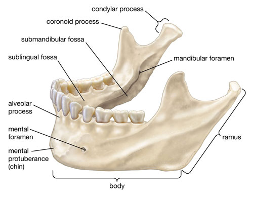
The mandible, more commonly known as the lower jawbone, is the largest and strongest bone in the human face. It supports the lower teeth and provides structure to the lower face. Playing a pivotal role in mastication (chewing), the mandible works in conjunction with the upper jaw, or maxilla, to process food and facilitate speech.
Maxilla
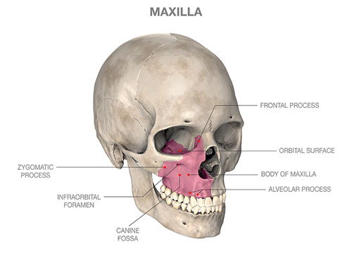
Opposite the mandible is the maxilla, or the upper jawbone. Apart from housing the upper teeth, the maxilla plays a key role in forming the roof of the mouth, the floor of the orbits (eye sockets), and the sides and floor of the nasal cavity. It’s crucial for functions like speaking and facial expressions.
Metacarpals
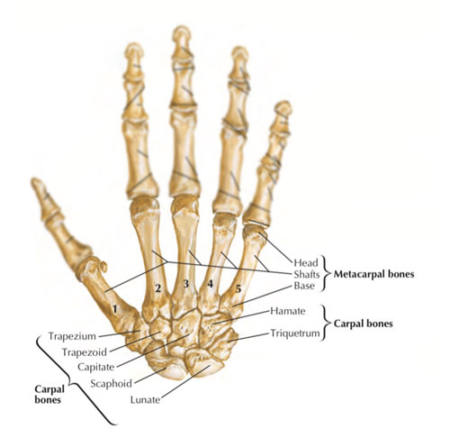
Moving on to the hands, the metacarpals are a group of five long bones situated in the palm. They are numbered from one to five, with one being adjacent to the thumb and five next to the little finger. These bones connect the wrist bones (carpals) to the finger bones (phalanges), aiding in hand flexibility and grip.
Metatarsals
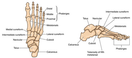
Similarly, in the feet, we have the metatarsals, which are a set of five long bones that play a role analogous to the metacarpals in the hand. They connect the ankle bones (tarsals) to the toes and are instrumental in walking, running, and bearing the body’s weight.
Meninges
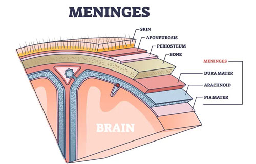
Diving deeper into the body, the meninges are three protective membranes that envelop the brain and spinal cord. These layers – the dura mater, arachnoid mater, and pia mater – safeguard the central nervous system, providing a barrier against physical impacts and infections.
Muscles
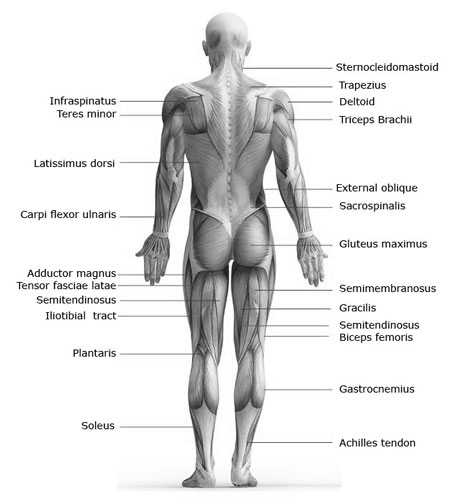
Perhaps the most extensive category under the letter “M” is muscles. Muscles are specialized tissues responsible for body movement, posture, and heat production. Ranging from the heart’s cardiac muscles to the biceps in our arms and the smooth muscles lining our internal organs, they are indispensable to nearly every bodily function.
Mammary Glands: More Than Just Breasts
The mammary glands, commonly known as breasts, are much more than just physical features. They play a vital role in nurturing life during motherhood. Composed of glandular tissue, fatty tissue, and connective tissue, these incredible organs have two main functions:
- Milk Production: During pregnancy and breastfeeding, the mammary glands produce milk to nourish newborns. Hormones signal the production of nutrient-rich breast milk, providing infants with all the essential minerals, vitamins, and antibodies they need to thrive.
- Emotional Connection: Breastfeeding fosters a powerful bond between mother and child, promoting emotional well-being and development. Skin-to-skin contact during feeding releases hormones that strengthen the maternal-infant relationship.
Did you know? The average woman has around 15-20 milk ducts in each breast, and each duct can have multiple branches! These tiny pathways transport milk from the milk-producing cells to the nipple for feeding.
Mandibular Nerve: The Mighty Mouth Mover
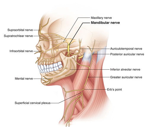
Imagine a complex electrical circuit controlling every bite, chew, and swallow. That’s the mandibular nerve, the largest branch of the trigeminal nerve, responsible for sensation and muscle movement in the lower jaw and face. Think of it as the master electrician running the show in your mouth and jaw!
- Sensational Savior: The mandibular nerve sends sensory information from the lower lip, chin, tongue, and teeth to the brain, allowing us to taste, feel temperature, and experience pain or pressure.
- Move It or Lose It: This mighty nerve also controls the muscles responsible for chewing, swallowing, and facial expressions. Without it, even simple actions like smiling or talking would be impossible!
Fun fact: The mandibular nerve is the only cranial nerve (nerves directly connected to the brain) with both sensory and motor functions. Talk about multitasking!
Maxillary Nerve: The Mischievous Maestro of the Mid-Face
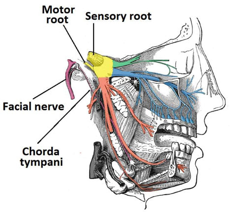
Meet the maxillary nerve, another branch of the trigeminal nerve, specializing in the upper half of your face. This mischievous maestro conducts a symphony of sensations and movements, keeping your face lively and expressive.
- Sensory Smorgasbord: The maxillary nerve delivers sensory information from the upper lip, cheek, nose, and eye to the brain, allowing you to feel pain, temperature, and touch. It also plays a crucial role in taste and smell.
- Facial Finesse: This nerve controls the muscles of the upper lip, eyelid, and nose, enabling you to blink, sniff, smile, and express a wide range of emotions.
Curious fact: The maxillary nerve supplies tear glands with the watery stuff for crying! So, next time you shed a tear, remember the mischievous maxillary nerve playing its part.
These are just glimpses into the remarkable world of the mammary glands, mandibular nerve, and maxillary nerve. As you explore these fascinating M-tastic features with your young learners, remember to emphasize the unique and vital roles they play in our bodies. And who knows, maybe these explorations will spark a lifelong love of learning about the incredible machinery that makes us human!
Median Nerve: The Mastermind of Movement
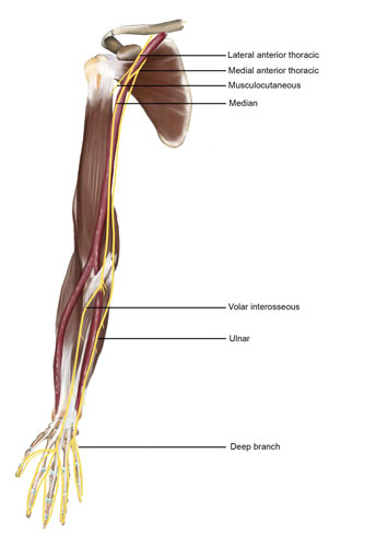
Imagine a highway running down your arm, carrying messages to and from your fingers. That’s the median nerve, a crucial player in sensation and movement in your upper limb. From the palm of your hand to the tip of your middle finger, this vital nerve is your connection to the world around you!
- Sensory Superstar: The median nerve collects sensory information from your fingers, palm, and forearm, sending it to your brain for interpretation. This allows you to feel touch, temperature, pain, and vibration, essential for everyday tasks like grasping objects or typing on a keyboard.
- Movement Maestro: This mighty nerve also controls the muscles in your forearm and hand, enabling you to flex, grip, pinch, and perform countless other dexterous movements. Without the median nerve, even simple actions like holding a spoon or writing your name would be impossible!
Did you know? Carpal tunnel syndrome, a common condition, occurs when the median nerve gets compressed in the wrist. Tingling, numbness, and weakness in the hand can be symptoms of this condition.
Mesentery: The Master Organizer of the Midsection
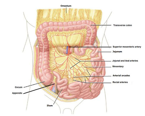
Hidden deep within your abdomen lies the mesentery, a thin, fold of tissue that holds your small intestine in place like a delicate hammock. But its role goes far beyond just being a gut-holding hero!
- Intestinal Architect: The mesentery anchors your small intestine, allowing it to move freely and efficiently as it digests food. It also supplies blood vessels, nerves, and lymphatics to the intestine, keeping it nourished and healthy.
- Drainage Director: This amazing fold also channels drainage from the intestine, moving waste products towards the lymphatic system and bloodstream for elimination.
Fun fact: The mesentery was only recently recognized as a distinct organ, with research suggesting it plays a crucial role in our immune system and overall gut health.
Mediastinum: The Middleman Maestro
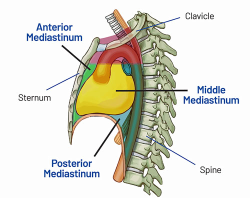
Think of your chest cavity as a bustling city, and the mediastinum as the central divider separating the “neighborhoods.” This sturdy wall of tissue, made of muscle and connective tissue, keeps everything organized and functioning smoothly.
- Master Divisor: The mediastinum separates the left and right sides of your chest, protecting the heart, lungs, and major blood vessels from bumping into each other. This ensures each organ has the space it needs to perform its vital tasks.
- Pathway Provider: This crucial dividing wall also provides passageways for vital structures like the esophagus, trachea, and major blood vessels, allowing them to connect different parts of your body effortlessly.
Did you know? The mediastinum contains the thymus gland, an essential part of the immune system that plays a vital role in early childhood development.
Midfoot: The Bridge Builder of the Foot
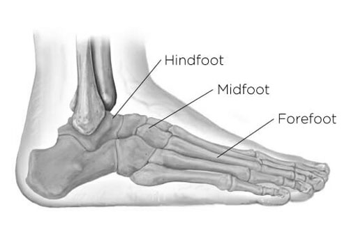
Nestled between the ankle and the forefoot lies the midfoot, a crucial bridge connecting the two halves of your foot. This sturdy platform isn’t just for show; it plays a vital role in stability, balance, and movement.
- Arch Architect: The midfoot houses the tarsal bones, which form the arches of your foot. These arches act like natural springs, absorbing shock and distributing pressure evenly when you walk, run, or jump.
- Balance Buddy: The midfoot helps maintain balance by connecting the ankle bones to the forefoot bones. This connection allows for subtle movements and adjustments that keep you upright and steady.
- Flexibility Facilitator: The midfoot also provides a flexible joint between the ankle and forefoot, enabling you to bend your foot and push off the ground for walking, running, and even dancing!
Did you know? The midfoot is particularly vulnerable to injuries like sprains and fractures due to its complex structure and weight-bearing role. Proper footwear and stretching can help prevent these problems and keep your midfoot strong and flexible.
Mouth: The Master of Munching and More
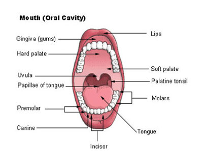
The mouth might seem like a simple gateway to food, but it’s a complex powerhouse packed with incredible features. From chomping to chirping, this mighty M-marvel plays a crucial role in our lives.
- Munching Maestro: The mouth’s starring cast includes 32 teeth (adults), designed to tear, grind, and chew food into digestible bits. Saliva, a watery concoction rich in enzymes, assists in breaking down food while tongue muscles perform a mesmerizing dance to mix and move everything around.
- Sensory Sentinel: The mouth is brimming with sensory receptors that help us taste sweet, salty, sour, bitter, and umami flavors. It also detects temperature and texture, allowing us to enjoy the full sensory experience of food.
- Communication Champion: Beyond just eating, the mouth is our tool for communication. Tongue movements and vocal cords work together to produce speech, allowing us to express ourselves, share stories, and connect with others.
Fun fact: The average human mouth produces around 2.5 liters of saliva every day! This amazing fluid not only aids digestion but also keeps the mouth clean and lubricated.
Muscular System: The Movers and Shakers
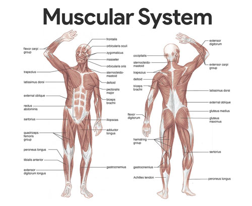
Imagine an orchestra where every instrument is a muscle, each playing a unique note to create the symphony of movement. That’s the muscular system, a complex network of over 600 muscles responsible for everything from blinking to sprinting.
- Movement Masters: Muscles work by contracting and relaxing, pulling bones and allowing for all kinds of motion. From walking and running to smiling and waving, the muscular system makes it possible for us to interact with the world around us.
- Posture Powerhouse: Core muscles in the back and abdomen provide stability and support, holding us upright and preventing slouching. Strong core muscles also improve balance and prevent back pain.
- Shape Shifters: Muscles can adapt and grow with training. Regular exercise helps build muscle mass and strength, improving overall health and performance.
Did you know? The smallest muscle in the human body is the stapedius, located in the middle ear and responsible for protecting the delicate inner ear structures from loud sounds.
Myocardium
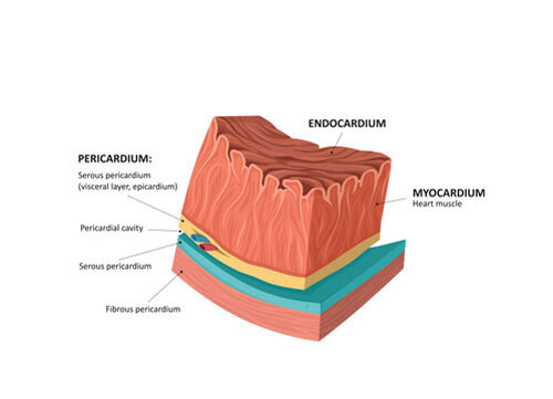
Speaking of muscles, the myocardium refers specifically to the heart muscle. This thick, muscular middle layer of the heart wall contracts rhythmically, pumping blood throughout the body. Any damage or ailment affecting the myocardium can have severe implications for cardiovascular health.
Malleus
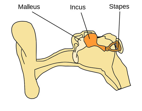
Within the confines of the ear lies the malleus, one of the three small ossicles in the middle ear. This hammer-shaped bone plays a crucial role in hearing by transmitting sound vibrations from the eardrum to the other ossicles and eventually to the inner ear.
Medulla Oblongata
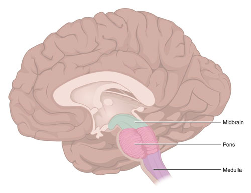
A component of the brainstem, the medulla oblongata, plays an essential role in regulating several autonomic functions, such as breathing, heart rate, and blood pressure. It’s the bridge between the brain and the spinal cord and is pivotal for maintaining basic life functions.
List of Human Body Parts Starting with M
| Male Genital Organs | Malleus | Mammary Glands |
| Mandibular Nerve | Masticatory Muscles | Maxillary Nerve |
| Maxillary Sinus | Medial Palpebral Ligament | Medial Pterygoid Plate |
| Medial Rectus Muscle | Median Nerve | Median Nerve, Motor Distribution |
| Mediastinum | Membranous Labyrinth | Meninges |
| Mesenteric Arteries | Mesentery | Metacarpophalangeal Joint |
| Middle Cerebral Artery | Middle Cranial Fossa | Midfoot |
| Mouth | Movements | Muscles |
| Muscles Of Breathing | Muscular System | Musculocutaneous Nerve |
| Mylohyoid Muscle | Maxilla | Mandible |
| Metacarpals | Metatarsals | Myocardium |
| Medulla Oblongata |
Conclusion
The letter “M” introduces us to a fascinating array of body parts, each integral to the mosaic of human life. From the bones that give our face structure to the muscles that enable movement, to the delicate components of our nervous system, the importance of these “M” parts is undeniable. This exploration underscores the marvel that is human anatomy, where every component, big or small, plays a role in the harmonious functioning of the body. As we continue our journey through the anatomy alphabet, we gain a deeper appreciation for the incredible machine that is the human body.
Human Body Parts That Start With
A | B | C | D | E | F | G | H | I | J | K | L | M | N | O | P | Q | R | S | T | U | V | W
Frequently Asked Questions
Are all 34 terms in the M list organs?
No. The directory mixes bones, nerves, muscles, membranes, spaces, regions, organs, and a body system. For example, the malleus is a small ear bone, the median nerve is a nerve, the mediastinum is a chest compartment, and the muscular system groups many muscles. Alphabetical lists are useful for vocabulary but do not imply that all entries are equivalent.
Is the maxillary nerve responsible for facial expressions?
No. The maxillary nerve (V2) is a sensory branch of the trigeminal nerve. It carries sensation from areas including the cheek, upper lip, upper teeth, palate, and lower eyelid. Facial-expression muscles are primarily controlled by the facial nerve (cranial nerve VII), while the mandibular branch of the trigeminal nerve supplies chewing muscles.
Does the median nerve supply the whole hand?
No. The median nerve supplies selected muscles and sensation to the palm side of the thumb, index and middle fingers, and thumb-side half of the ring finger, with some additional fingertip territory. The ulnar and radial nerves serve other parts of the hand. The median nerve can be compressed in the carpal tunnel at the wrist.
Is the mesentery a newly discovered organ?
No. The mesentery has been described in anatomy for centuries. Recent discussion has emphasized its continuous structure and prompted debate about its classification as an organ. In practical terms, it is a peritoneal fold that supports parts of the intestine and carries blood vessels, nerves, and lymphatic vessels.
Where is the medulla oblongata?
The medulla oblongata is the lowest portion of the brainstem, where it continues into the spinal cord. It contains nerve pathways and centers involved in automatic functions including breathing and cardiovascular regulation. Describing it only as a bridge misses its active role in processing and regulating signals.
How many teeth and how much saliva does an adult usually have?
Adults often have 28 teeth without wisdom teeth or up to 32 when all third molars are present. Saliva production varies by person and conditions; Cleveland Clinic reports about 0.5 to 1.5 liters daily for most adults. These figures are ranges, not exact amounts that apply to everyone.
Sources and further reading
- NCBI Bookshelf: The Meninges — protective layers around the brain and spinal cord.
- NCBI Bookshelf: Medulla Oblongata — brainstem anatomy and function.
- NCBI Bookshelf: Anatomy, Thorax — mediastinum and thoracic compartments.
- NCBI Bookshelf: Trigeminal Neuropathy — V2 and V3 sensory and motor distributions.
- NCBI Bookshelf: Median Nerve — forearm, hand, and sensory distributions.
- NCBI Bookshelf: Small Intestine and Peritoneum — mesentery and its connections.
- Cleveland Clinic: Ossicles — sound transmission through the malleus, incus, and stapes.
- Cleveland Clinic: Saliva — typical adult saliva production range.