In the expansive lexicon of human anatomy, each letter unfolds a unique chapter, brimming with its own set of tales and intricacies. As we journey alphabetically, some letters, such as ‘U’, might seem to hold fewer entries, but their significance in the overarching narrative of our physiology is profound.
This article sets the stage for a deep dive into the ‘U’-initiated body components, parts that may often be overshadowed by more frequently discussed anatomical structures. From the ubiquitous utility of our ulna to the unsung undertakings of the urethra, we will explore the nuances, roles, and interplay of these essential components. Prepare to venture into the less charted territories of the human body, unraveling the mysteries and marvels that commence with the letter “U.”
Human Body Parts That Start With The Letter U
Our journey through the alphabet of human anatomy has offered insights into the beauty, complexity, and efficiency of the body’s many components. As we reach the letter “U,” we enter a more compact domain, characterized by specific yet significant structures. Although fewer in number, the body parts beginning with “U” play crucial roles in various physiological processes. Let’s delve into these parts, shedding light on their function, significance, and intricacies.
Ulna
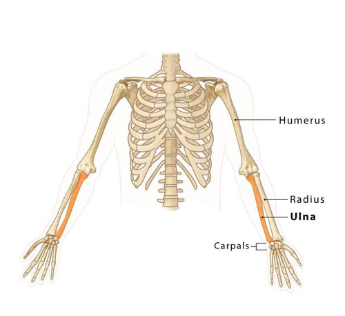
Found in the forearm, the ulna is one of the two long bones (with the other being the radius) that stretch from the elbow to the wrist. The ulna is larger and situated medially when the hand faces forward. It is instrumental in forming the elbow joint with the humerus of the upper arm. The ulnar bone’s distinctive “U” shape at its top end provides a hinge for the elbow’s flexion and extension.
Urethra
The urethra is a tubular structure responsible for transporting urine from the bladder to the outside of the body. Its role varies slightly between males and females. In males, the urethra also serves as a conduit for semen during ejaculation, while in females, its primary function is urine excretion.
Uterus
An essential component of the female reproductive system, the uterus or womb is a muscular organ where a fertilized egg implants and grows during pregnancy. The uterus consists of the fundus (upper part), corpus (body), and cervix (lower part). It undergoes significant changes during a woman’s menstrual cycle and pregnancy, highlighting its dynamic nature.
Uvula
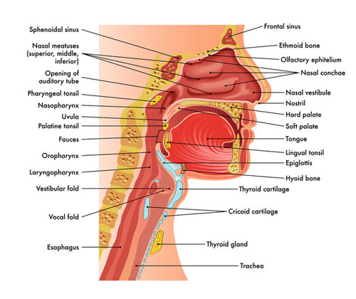
Located at the back of the throat and dangling from the soft palate, the uvula is a small, fleshy extension. While its exact purpose isn’t entirely understood, it’s believed to assist in speech production and prevent food from entering the nasal passage.
Ulnar Artery
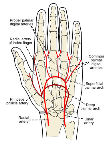
A significant blood vessel of the forearm, the ulnar artery runs alongside the ulna bone. It arises from the brachial artery and provides oxygenated blood to the medial aspects of the forearm, eventually participating in the formation of the superficial and deep palmar arches in the hand.
The Unsung Hero of Your Hand: The Ulnar Nerve
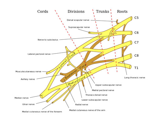
Have you ever stopped to consider how you effortlessly pinch, grasp, and tickle? It’s all thanks to the ulnar nerve, a hidden champion working tirelessly within your arm and hand. This mighty nerve, nicknamed the “musician’s nerve”, plays a crucial role in your hand’s fine motor skills.
Imagine the ulnar nerve as your hand’s personal electrician, sending vital nerve signals to various muscles. It controls the flexing and spreading of your pinkie and ring finger, allowing you to hold a pen, grab a ball, or play a musical instrument with precision. Did you know that nearly 50% of the hand’s muscles rely on the ulnar nerve for movement? That’s right, almost half your hand’s dexterity hinges on this single nerve!
But the ulnar nerve isn’t just a mover and shaker. It’s also a skilled information gatherer. Its sensory branches provide feeling to the little finger and half of the ring finger, both on the palm and back sides. This allows you to distinguish a soft touch from a rough scratch, even on these tiny digits. This sensory feedback is crucial for everyday tasks like buttoning your shirt or feeling the texture of different fruits.
So next time you admire your hand’s amazing skills, remember the silent conductor behind the scenes – the ulnar nerve. This unsung hero deserves a round of applause for keeping your hand nimble and your senses sharp!
Building Bridges and Tunnels: The Bones of Your Upper Limbs
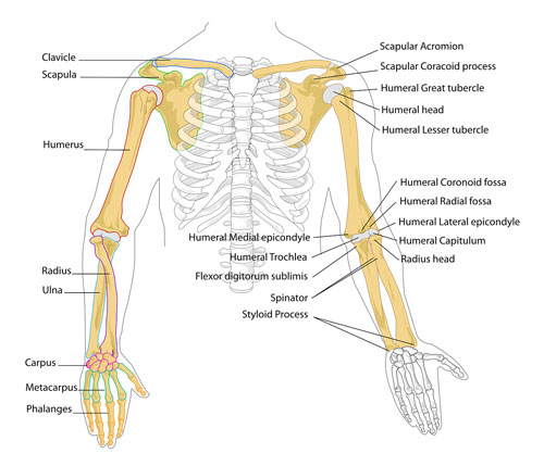
Your arms and hands aren’t just waving and grabbing machines; they’re engineering marvels built on a strong foundation – the bones of your upper extremities. These 30 skeletal wonders, from shoulder to fingertips, work together to provide support, movement, and protection for all your actions, big and small.
Let’s start at the base. The shoulder girdle forms the bridge connecting your arm to your body. Two main bones hold things together: the clavicle, a slender collarbone like a suspension bridge, and the scapula, a wing-shaped shoulder blade providing anchor points for muscles.
Next, meet the humerus, the single sturdy bone of your upper arm. Think of it as a highway tunnel, carrying nerves and blood vessels alongside powerful muscles. At the elbow, the tunnel splits into two lanes: the radius on the thumb side and the ulna on the pinky side. These forearm bones work together with a hinge-like joint, allowing you to bend and straighten your arm like a drawbridge.
Finally, we reach the bustling city of your hand. Here, 27 small bones, the carpus (wrist), metacarpals (palm), and phalanges (fingers and thumb), come together like intricate bridges and pathways. This complex network allows for the incredible flexibility and precision of your fingers, letting you write, play instruments, and do countless other delicate tasks.
So, the next time you reach out to grab a toy, wave hello, or build a sandcastle, remember the amazing team effort happening inside your arm. From the sturdy tunnels of the arm bones to the bustling metropolis of your hand, your upper extremity bones are the tireless engineers behind every action!
The Silent Rivers of Your Arms: Upper Extremity Veins
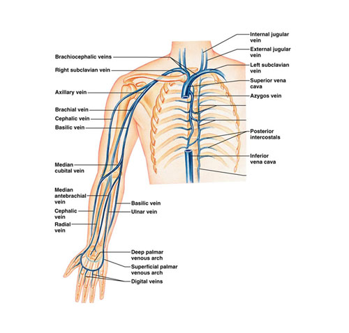
While arteries get all the credit for delivering oxygen-rich blood to your arms, another crucial set of vessels quietly carries the used blood back – the upper extremity veins. Though less glamorous than their arterial counterparts, these silent heroes play a vital role in keeping your arms and hands healthy and functional.
Imagine your upper extremity veins as a network of underground rivers, flowing back towards your heart. Similar to arteries, this network has two main systems: the superficial and deep. The superficial veins, like the easily visible blue lines on your forearms and hands, lie close to the skin’s surface. They’re responsible for draining blood from the muscles and skin. Meanwhile, the deep veins, located deeper within the muscles and alongside arteries, carry the majority of the blood back to the heart.
These upper extremity veins aren’t simply passive pipelines. They have built-in valves that prevent blood from flowing back down, ensuring a one-way trip towards the heart. These valves are especially important in the legs, where gravity helps pull blood against the flow. In the arms, muscle contractions help squeeze the veins and push blood uphill.
So, the next time you raise your arm to wave or reach for a treat, remember the silent rivers flowing within. These upper extremity veins are vital partners in the continuous cycle of blood circulation, keeping your arms and hands functioning flawlessly. They’re a testament to the body’s amazing design, where every part, even the less flashy ones, plays a crucial role in keeping us healthy and alive.
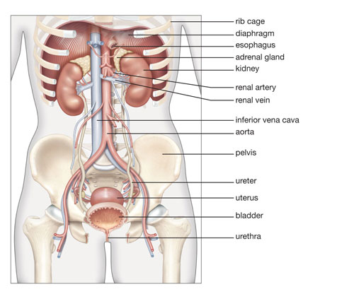
The Unsung Heroes of Pee: The Ureters
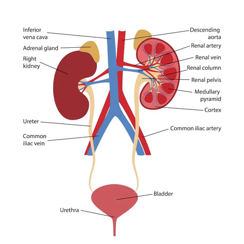
We all know about the kidneys, those busy bean-shaped organs that filter our blood and create urine. But what happens to that urine once it’s made? That’s where the ureters come in, two unsung heroes silently whisking your waste away.
Imagine the ureters as tiny, muscular water slides, about as thick as a drinking straw and roughly 25 centimeters long. Each starts at the bottom of a kidney, like a drain plug, and winds its way down through your abdomen and pelvis until it reaches the bladder. That’s right, they snake all the way from your back to your lower belly!
These little tubes aren’t just passive pipes, though. They’re lined with smooth muscle that contracts in waves, propelling urine downwards like a miniature water pump. This rhythmic movement, called peristalsis, happens about three times a minute, ensuring a steady flow without needing any conscious effort.
So next time you take a bathroom break, give a silent cheer for the amazing ureters. These tireless tubes work 24/7, quietly taking care of the dirty work to keep you feeling healthy and comfortable. They’re a true testament to the body’s intricate design, where even the smallest parts play a vital role in our well-being.
Bonus fact: Did you know that ureters are surprisingly tough? They can stretch and twist to accommodate our movements without getting damaged. That’s why they can handle even the most energetic jumps and tumbles!
The Tiny Gatekeeper: The Urethral Sphincter Muscle
Tucked away within the pelvic floor lies a tiny but mighty muscle called the urethral sphincter. Don’t let its size fool you; this vital muscle plays a crucial role in one of our most basic functions – controlling urine flow.
Imagine the urethral sphincter as a flexible drawstring wrapped around the urethra, the tube that carries urine from the bladder to the outside world. When relaxed, the drawstring loosens, allowing urine to flow freely. But when we need to hold it, the sphincter muscle contracts, tightening the drawstring and blocking the flow of urine. Think of it as a tiny traffic light, switching between green (go) and red (stop) for urine flow.
This remarkable muscle works in two parts:
- Internal urethral sphincter: This involuntary muscle, controlled by the nervous system, is constantly active, maintaining a baseline level of closure even when we’re not consciously thinking about it. It’s like an automatic door closer for the urethra, ensuring we don’t have accidental leaks.
- External urethral sphincter: This voluntary muscle is the one we actively control when we need to hold or release urine. By squeezing or relaxing this muscle, we can open or close the “gate” to the urethra, allowing us to control urination.
The urethral sphincter is essential for maintaining urinary continence, meaning we can control when and where we release urine. It’s especially important for young children who are still learning to potty train, as well as for adults who may experience occasional leaks due to medical conditions or aging.
So next time you take a trip to the bathroom, remember the incredible urethral sphincter muscle working quietly behind the scenes. This tiny gatekeeper plays a vital role in keeping us dry and comfortable, allowing us to go about our daily lives without worry.
The Amazing Journey: Exploring Your Urinary Tract
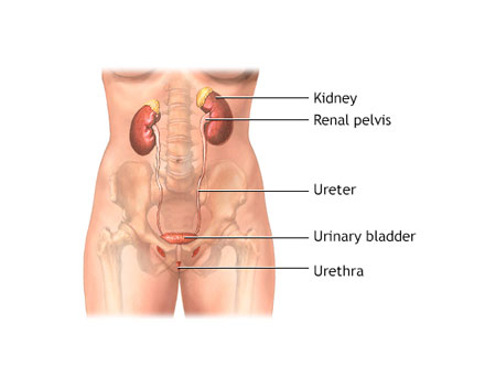
Imagine a bustling highway system within your body, constantly filtering waste and keeping things running smoothly. That’s exactly what the urinary tract does! This incredible network of organs works tirelessly to remove waste products from your blood, produce urine, and send it out of your body. Let’s take a closer look at this fascinating system:
The Filtration Powerhouse:
- Kidneys: These bean-shaped organs, located near your back, are the stars of the show. They act as filtration units, constantly processing your blood and separating waste products like urea and excess water. Each day, your kidneys filter about 150 liters of blood, producing around 1-2 liters of urine!
Kidneys in the human body
The Downhill Delivery:
- Ureters: Think of these as two thin tubes, each connecting a kidney to the bladder. They carry the newly formed urine downhill from the kidneys to the bladder, using muscular contractions to keep the flow going.
Ureters in the human body
The Storage Hub:
- Bladder: This muscular sac acts as a reservoir for urine. As the ureters deliver urine, the bladder stretches to accommodate it, allowing you to go about your day without needing to constantly use the bathroom. The healthy adult bladder can typically hold around 400-600 milliliters of urine before feeling the urge to empty.Opens in a new windowmy.clevelandclinic.orgBladder in the human body
The Exit Gate:
- Urethra: This tube connects the bladder to the outside of your body. When you’re ready to pee, the bladder muscles contract, pushing urine out through the urethra. A sphincter muscle around the urethra controls the flow, allowing you to hold it when needed.
Fun Facts for Kids:
- Did you know your kidneys are about the size of your fists?
- Urine is mostly water, but it also contains important chemicals like urea and creatinine.
- The color of your urine can tell you a lot about your health. Pale yellow is ideal, while dark yellow or reddish hues can indicate dehydration or other issues.
- It takes about 40 seconds for urine to travel from your kidneys to your bladder!
Remember:
The urinary tract is a vital part of your body, keeping you healthy and comfortable. Taking care of it is important! Drink plenty of water, avoid holding your pee for too long, and practice good hygiene to keep your urinary tract happy and healthy.
The Hidden Highway to Miracles: The Uterine Tubes
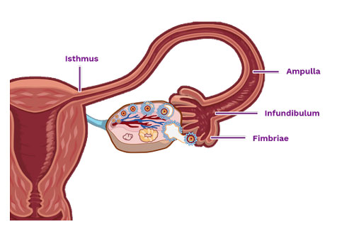
Imagine a pair of delicate, shimmering tubes tucked away safely within your abdomen, each no thicker than a strand of spaghetti. These aren’t just any ordinary tubes; they’re the uterine tubes, also known as fallopian tubes, and they play a starring role in the incredible journey of life!
Think of the uterine tubes as secret highways connecting your ovaries, where eggs are made, to your uterus, where babies grow. Each tube is about 10-12 cm long and lined with tiny hair-like structures called cilia. These cilia wave back and forth like tiny flags, creating a current that sweeps any mature egg released from the ovary down the tube towards the uterus.
Uterine tubes in the female reproductive system
But the uterine tubes aren’t just passive pathways. They’re like bustling city streets filled with activity! Inside them, a special fluid nourishes the egg and provides a welcoming environment for something truly magical to happen – fertilization! If a sperm happens to meet the egg on its journey, they undergo a dance called fertilization, merging their genetic material to create the very first cell of a new life.
Once fertilized, the egg, now an embryo, continues its journey down the tube, propelled by the cilia and muscular contractions. It takes about 3-5 days for the tiny embryo to reach the cozy haven of the uterus, where it implants and begins its incredible transformation into a baby.
So next time you marvel at the miracle of life, remember the unsung heroes behind the scenes – the uterine tubes. These incredible little highways, with their tireless cilia and welcoming fluid, pave the way for every new beginning, making them one of the most important body parts in the story of life.
Fun facts for kids:
- Did you know the uterine tubes are lined with millions of cilia? That’s a lot of tiny flags working together!
- An egg is only about the size of a grain of sand, but it holds the potential for a whole new life!
- Fertilization usually happens in the outer third of the fallopian tube, closer to the ovary.
Remember:
The uterine tubes are essential for healthy reproduction. Taking care of your body by eating healthy, staying active, and getting regular checkups helps keep them healthy too!
Umbilicus
Commonly known as the navel or belly button, the umbilicus is a central scar on the abdomen. It is the remnant of where the umbilical cord attached a fetus to its mother’s placenta, facilitating the transfer of nutrients, oxygen, and waste products.
Uvea
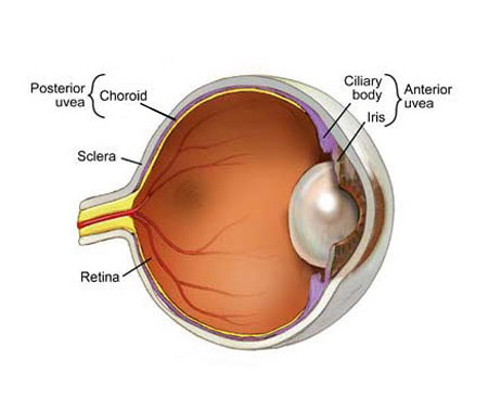
A critical layer of the eye, the uvea lies beneath the white sclera and consists of three main parts: the iris (colored part of the eye), ciliary body (a muscle that helps the lens focus), and choroid (a layer of blood vessels and connective tissues). The uvea supplies nutrients to various parts of the eye and is instrumental in vision.
List of Human Body Parts Starting with U
| Ulnar Artery | Ulnar Nerve | Ulnar Nerve, Motor Distribution |
| Upper Extremity Bones | Upper Extremity Veins | Ureter |
| Urethra | Urethral Sphincter Muscle | Urinary Tract |
| Uterine Tube | Uterus | Uvea |
| Umbilicus | Uvula | Ulnar |
| Upper Limbs |
Conclusion
The realm of “U” in human anatomy might be succinct, but its components are undeniable in their importance. From the strength-bearing ulna of our forearm to the nutrient-providing uvea of our eyes, each structure emphasizes the meticulous design and function of our body. While some of these parts might be less discussed in common parlance, their roles in maintaining our physiological balance cannot be understated. This exploration into the “U” components of our anatomy serves as a testament to the idea that no matter how seemingly minor a part may appear, each contributes profoundly to the symphony of human existence.
Human Body Parts That Start With
A | B | C | D | E | F | G | H | I | J | K | L | M | N | O | P | Q | R | S | T | U | V | W