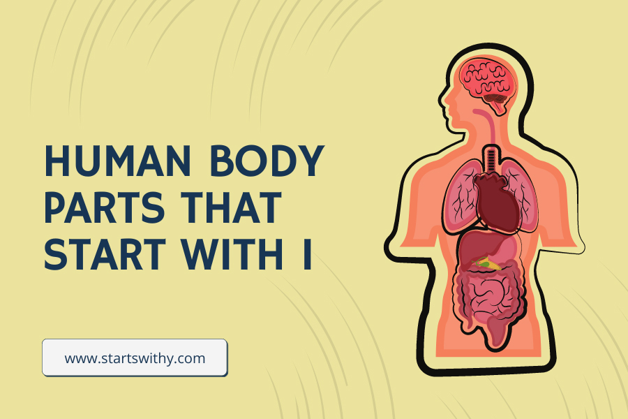
Immerse yourself in the intricate world of human anatomy with our latest article, “Body Parts That Start With I”. This enlightening exploration focuses on those parts of our bodies that start with the letter ‘I’, from the integral organ, the intestines, to the microscopic insulin-producing cells. We delve into their structure, function, and unique characteristics, providing an engaging and informative read.
This piece is an excellent resource for students, educators, or anyone with an interest in the miraculous human body. Whether you’re a seasoned medical professional or a novice in the field of biology, join us as we continue our alphabetical journey through the stunning complexity of our bodies.
Human Body Parts That Start With The Letter I
The human body is an intricate and remarkable system, made up of numerous parts, each contributing to our overall health and functioning. Continuing our alphabetical exploration of the human body, we’ve arrived at the letter “I.” This letter may not represent a large quantity of body parts, but the ones it does designate are essential to our wellbeing. This article aims to delve into the details of these body parts that start with “I,” discussing their structure, function, and their roles in our health.
Ilium
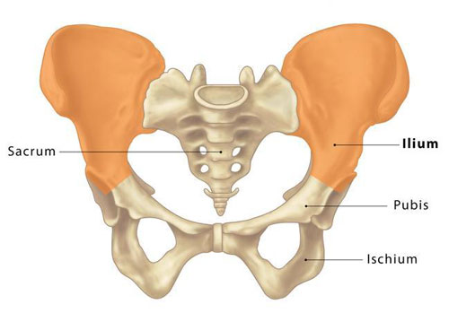
The ilium is the largest and most superior of the three bones that make up the human pelvis (the other two being the ischium and the pubis). The ilium forms the broad, flared portion of the pelvis, known as the hip bone. It plays a crucial role in supporting the body’s weight and providing attachment points for several muscles, including those of the lower back, abdomen, and hip.
Iris
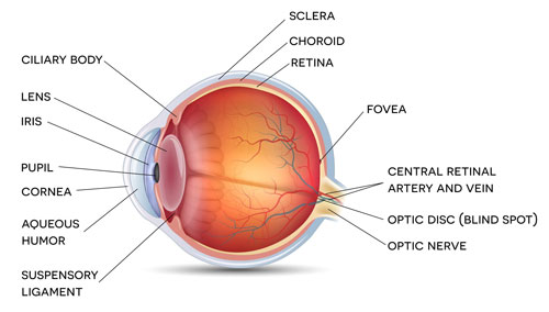
The iris is the circular, pigmented membrane that provides our eyes with their distinctive color. Located behind the cornea and in front of the lens, the iris has an adjustable circular opening in its center called the pupil. The iris’s primary function is to control the diameter and size of the pupil, thereby regulating the amount of light that reaches the retina, much like the aperture of a camera.
Intestines
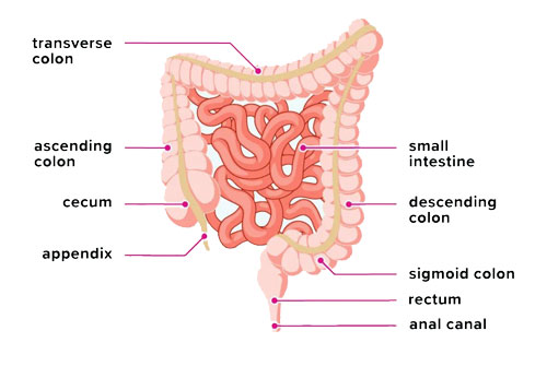
The intestines, a critical component of the digestive system, are a long, continuous tube running from the stomach to the anus. They are divided into two main sections: the small intestine and the large intestine. The small intestine’s role involves the digestion and absorption of nutrients, while the large intestine primarily absorbs water and forms feces.
Iliotibial Band
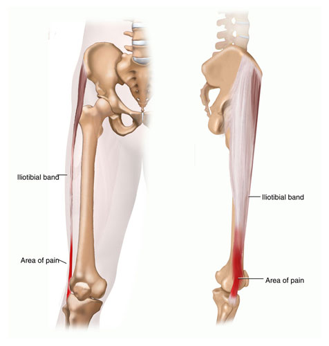
The iliotibial band, often referred to as the IT band, is a long piece of connective tissue, or fascia, that runs along the outside of the thigh. It starts at the ilium of the pelvis, crosses the hip and knee, and attaches to the top of the tibia (shin bone). The IT band helps stabilize the knee and assists in movements such as walking and running.
Incus
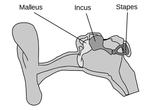
The incus is one of the three tiny bones, collectively known as the ossicles, located in the middle ear. These three bones—the malleus, incus, and stapes—work together to transmit and amplify sound waves from the outer ear to the inner ear. The incus, specifically, receives vibrations from the malleus, to which it’s connected, and passes them onto the stapes.
Inferior Vena Cava
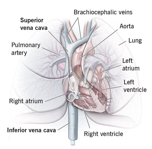
The inferior vena cava (IVC) is the largest vein in the body, carrying deoxygenated blood from the lower half of the body back to the heart. It starts from the convergence of the two major veins from the legs and ends at the right atrium of the heart. The blood returned by the IVC is then pumped by the heart to the lungs to be oxygenated.
Intercostal Muscles
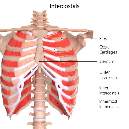
The intercostal muscles are several groups of muscles that run between the ribs, helping form and move the chest wall. These muscles are chiefly involved in the mechanical aspect of breathing, aiding in the expansion and contraction of the chest cavity during respiration.
Iliac Artery: The Blood Supply Superhighway
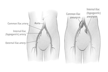
Imagine a mighty river branching out into smaller tributaries – that’s the iliac artery, the main blood vessel supplying the lower body. This vital highway carries oxygen-rich blood from the aorta, the body’s main artery, to the pelvic region, legs, and feet.
- Branching Out: The iliac artery splits into two main branches just below the belly button – the common iliac arteries. These arteries then further divide into the internal and external iliac arteries, delivering blood to specific regions:
- Internal Iliac Artery: This branch supplies blood to the pelvic organs, bladder, and internal reproductive organs.
- External Iliac Artery: This branch travels down the leg, eventually becoming the femoral artery, the main blood vessel supplying the thigh and leg.
- Powering Movement: The iliac artery plays a crucial role in movement. During exercise, the artery dilates, allowing more blood to flow to the legs, providing the muscles with the oxygen they need to keep us moving.
- Maintaining Health: Blockages or narrowing of the iliac artery can lead to serious health problems like leg pain, reduced mobility, and even tissue death. Regular exercise, a healthy diet, and managing risk factors like diabetes and high blood pressure are crucial for keeping these vital pathways healthy.
Iliac Vein: The Return Route
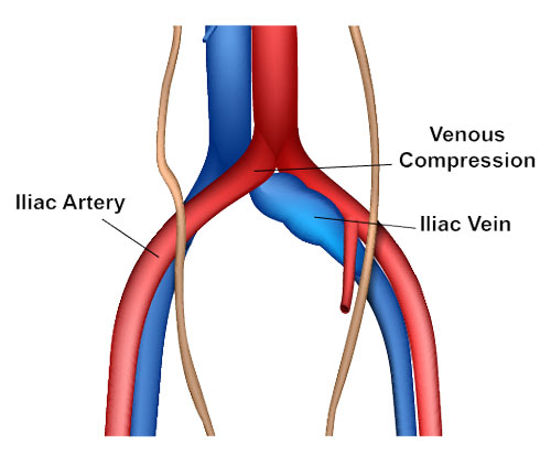
While the iliac artery delivers fresh blood, the iliac vein acts like a return route, carrying deoxygenated blood from the lower body back to the heart. Imagine it as the river tributaries flowing back into the main stream.
- Dual Drainage: Similar to the iliac artery, the iliac vein also has two main branches – the common iliac veins. These veins then merge into the inferior vena cava, the largest vein in the body, which carries blood back to the heart.
- Waste Removal: The iliac vein carries waste products like carbon dioxide and lactic acid produced by the muscles back to the lungs and liver for processing and elimination.
- Maintaining Pressure: Valves within the iliac vein prevent blood from flowing backward, ensuring efficient drainage and proper blood pressure in the lower body.
Inner Ear: The Balance and Hearing Maestro
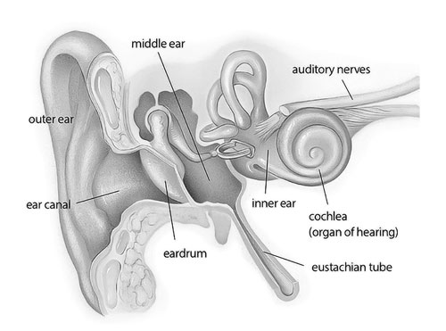
Hidden deep within the skull lies the inner ear, a marvel of engineering responsible for our sense of balance and hearing. This tiny, fluid-filled labyrinth houses delicate structures that translate sound waves and head movements into electrical signals sent to the brain.
- Balance Champions: The semicircular canals and otoliths within the inner ear detect changes in head position and movement. These tiny structures send signals to the brain, allowing us to maintain balance, coordinate our movements, and prevent dizziness.
- Hearing Heroes: The cochlea, another part of the inner ear, is shaped like a snail shell and houses hair cells that respond to sound vibrations. These vibrations are converted into electrical signals that travel to the brain, allowing us to hear and understand sounds.
- Protecting the Masterpiece: The inner ear is a delicate and sensitive organ. Loud noises, ear infections, and certain medications can damage its structures, leading to hearing loss or balance problems. Protecting our ears from loud noises and maintaining good ear hygiene are crucial for keeping this vital conductor in tune.
Inguinal Canal: The Gateway to Growth
Tucked within the lower abdomen, nestled between the thigh and belly, lies the inguinal canal. This narrow passageway acts as a crucial route for vital structures, playing a vital role in development and function.
Inguinal canal showing its location and surrounding structures
- Life-Giving Passage: During fetal development, the inguinal canal serves as a pathway for the testicles in males and ovaries in females to descend from the abdomen into the groin. This migration is essential for proper reproductive function.
- Muscle Maestro: The canal is reinforced by strong muscles like the external oblique and transversus abdominis. These muscles contract during activities like coughing or lifting, creating pressure that helps prevent hernias, bulges of abdominal tissue through the weak points in the canal wall.
- Surgical Significance: Understanding the anatomy of the inguinal canal is crucial for surgeons performing hernia repairs. They carefully access the canal, manipulate the protruding tissue back into the abdomen, and strengthen the weak spots to prevent future hernias.
Inspiration: The Breath of Life
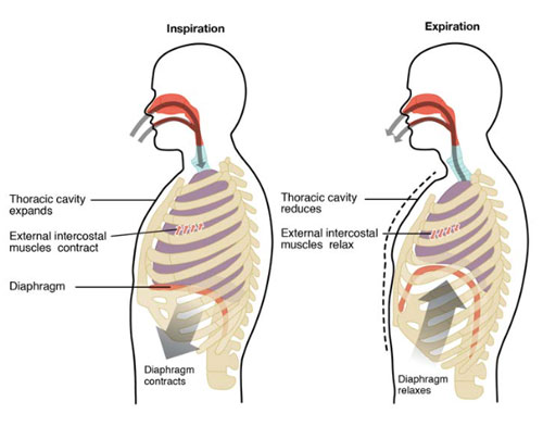
Have you ever stopped to appreciate the miracle of taking a breath? This seemingly effortless act involves a complex symphony of muscles, nerves, and air passages, culminating in the wonder of inspiration.
- Diaphragm Dynamo: The star of the show is the diaphragm, a dome-shaped muscle located below the lungs. When it contracts, it flattens, creating a vacuum in the chest cavity, drawing air into the lungs through the trachea.
- Intercostal Ensemble: Between the ribs lie the intercostal muscles, which contribute to breathing by expanding and contracting the rib cage. Their coordinated movement with the diaphragm ensures smooth and efficient air intake.
- Nerve Network: The entire operation is orchestrated by a network of nerves, including the phrenic nerve, which controls the diaphragm, and the intercostal nerves, which control the intercostal muscles. These nerves send signals from the brain, regulating the rhythm and depth of our breaths.
Intercostal Nerves: The Messengers Between Mind and Muscle
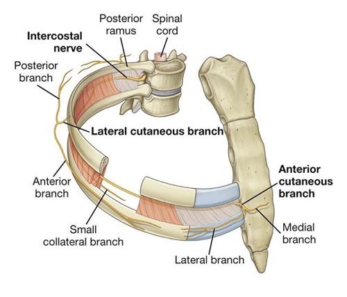
Weaving their way through the rib cage, nestled between the intercostal muscles, lie the intercostal nerves. These vital messengers play a crucial role in movement, sensation, and even reflexes.
- Motor Marvels: The intercostal nerves carry signals from the spinal cord to the intercostal muscles, controlling their contraction and relaxation. This enables us to cough, sneeze, breathe deeply, and perform various movements involving the chest and upper back.
- Sensory Spies: These nerves also transmit sensory information from the skin, muscles, and organs of the chest and abdomen back to the brain. This allows us to feel pain, temperature, pressure, and even the movement of our internal organs.
- Reflex Champions: Some intercostal nerves are involved in important reflexes like the cough reflex, which helps clear the airways of irritants, and the hiccup reflex, which is an involuntary contraction of the diaphragm.
Ischium: The Seat of Strength
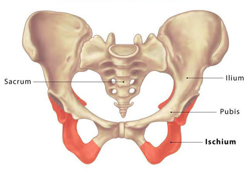
Imagine a sturdy, bony platform forming the base of your pelvis – that’s the ischium. This unsung hero provides support for sitting, walking, and various other movements, making it a vital part of our musculoskeletal system.
- Powerhouse for Sitting: The ischium, along with the ilium and pubis, forms the sit bone. This sturdy structure provides a stable base for sitting, absorbing pressure and distributing our weight evenly.
- Muscle Magnet: The ischium serves as an attachment point for several key muscles, including the hamstrings, gluteus maximus, and adductors. These muscles work together to power walking, running, and other movements of the legs and hips.
- Injury Prevention: Strong muscles surrounding the ischium can help prevent injuries like bursitis and tendinitis, which can cause pain and discomfort in the sitting and hip area.
Intervertebral Discs: Cushioning the Spine
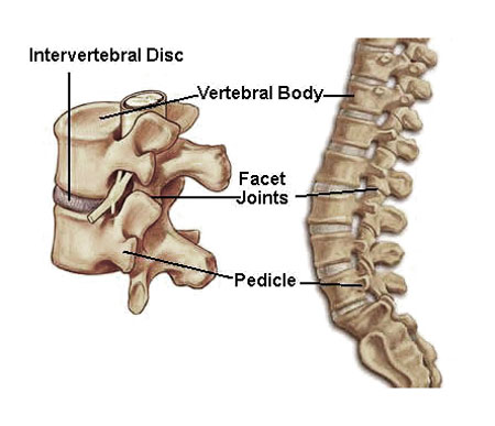
Imagine a series of soft, jelly-like cushions nestled between your vertebrae – that’s the intervertebral discs. These crucial structures act as shock absorbers, protecting the spine from impact and providing flexibility for movement.
- Spinal Support System: Each disc is composed of a tough outer ring (annulus fibrosus) and a soft inner core (nucleus pulposus). This combination provides cushioning and distributes pressure evenly across the spine.
- Movement Magic: Intervertebral discs allow for a remarkable range of motion in the spine, enabling us to bend, twist, and turn with ease. They also play a crucial role in maintaining good posture and absorbing shock during activities like jumping or running.
- Disc Degeneration: Over time, intervertebral discs can wear and tear, leading to conditions like herniation or bulging. Maintaining good posture, strengthening core muscles, and avoiding heavy lifting can help prevent disc problems and keep your spine healthy.
Innominate Artery: The Blood Supply Highway
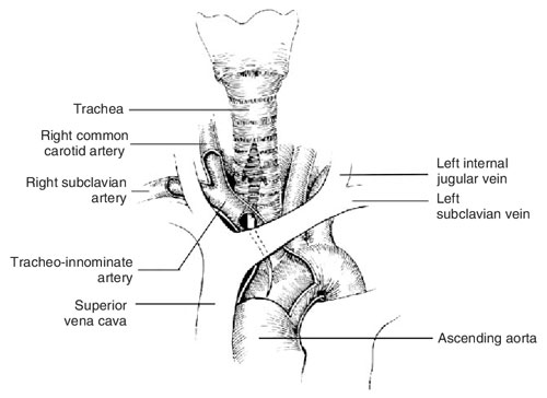
Imagine a mighty river branching out into smaller tributaries – that’s the innominate artery, the main blood vessel supplying the upper body. This vital highway carries oxygen-rich blood from the aorta, the body’s main artery, to the head, neck, arms, and chest.
- Branching Out: The innominate artery divides into two main branches just above the collarbone – the left and right subclavian arteries. These arteries then further branch out to supply blood to specific regions:
- Left Subclavian Artery: Supplies blood to the left arm, shoulder, and head.
- Right Subclavian Artery: Supplies blood to the right arm, shoulder, and head.
- Fueling Movement: The innominate artery plays a crucial role in providing blood to the muscles during exercise. During physical activity, the artery dilates, allowing more blood to flow to the arms and head, providing them with the oxygen they need to keep us moving.
- Maintaining Health: Blockages or narrowing of the innominate artery can lead to serious health problems like stroke, heart attack, and even vision loss. Regular exercise, a healthy diet, and managing risk factors like high blood pressure and cholesterol are crucial for keeping this vital pathway healthy.
Islets of Langerhans
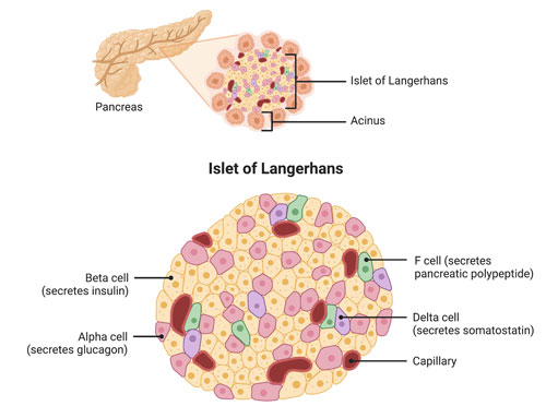
The Islets of Langerhans, often simply referred to as pancreatic islets, are tiny clusters of cells scattered throughout the pancreas. These cells play a vital role in endocrine function. They are responsible for the production and release of hormones such as insulin (which lowers blood sugar levels) and glucagon (which raises blood sugar levels), playing a vital role in maintaining glucose homeostasis in the body.
List of Human Body Parts Starting with I
| Iliac Artery | Iliac Vein | Incus |
| Inferior Alveolar Nerve | Inferior Cerebellar Arteries | Inferior Epigastric Artery |
| Inferior Oblique Muscle | Inferior Rectus Muscle | Inferior Vena Cava |
| Inguinal Canal | Inguinal Ligament | Inner Ear |
| Innominate Artery | Inspiration | Inter-Atrial Septum |
| Intercostal Nerves | Internal Carotid Artery | Interstitium |
| Interventricular Septum | Intervertebral | Intervertebral Foramen |
| Intestines | Ischio-Cavernosus Muscle | Ischium |
| Ilium | Iris | Iliotibial Band |
| Intercostal Muscles | Islets of Langerhans |
Conclusion
The exploration of body parts beginning with “I” reveals a fascinating mixture of structures, each playing a critical role in our health and functioning. From the ilium, a significant component of our skeletal system, to the islets of Langerhans, playing a key role in endocrine regulation, these “I” body parts showcase the intricacies and wonder of human anatomy. This exploration reminds us that every piece of the human body, no matter how small, is vital to the remarkable machine that keeps us alive and functioning every day.
Human Body Parts That Start With
A | B | C | D | E | F | G | H | I | J | K | L | M | N | O | P | Q | R | S | T | U | V | W
