How to read this D anatomy list
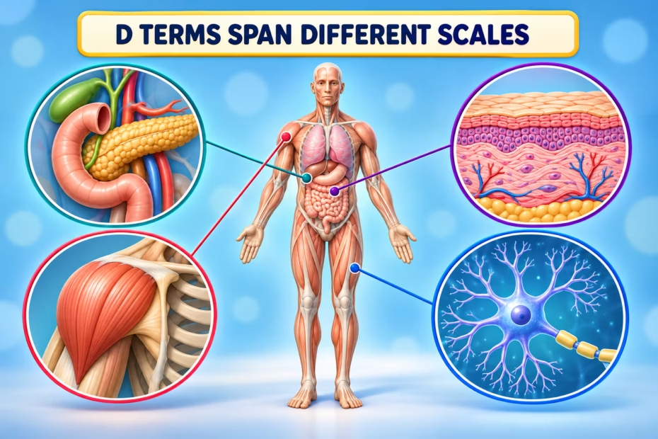
The original directory contains 16 entries, but they do not all name the same kind of thing. It includes organs and tissues, muscles, nerves, blood vessels, cellular structures, DNA, and the general term ductus. These are useful vocabulary examples, rather than 16 separate organs. The notes below clarify several locations and functions; the complete original article follows unchanged.
| Kind of structure | Examples from the D list | What the term describes |
|---|---|---|
| Organs, tissues, and regions | Duodenum, dermis, dental pulp, diaphragm | Parts of the digestive tract, skin, tooth, and muscle partition between chest and abdomen. |
| Muscles, nerves, and neuron parts | Deltoids, digital nerves, dendrites | Structures that produce movement or carry and receive nervous-system signals. |
| Vessels, membranes, and channels | Descending aorta, dura, ducts, ductus | Terms with specific anatomic locations; “ductus” alone is a broad Latin-derived term for a passage or duct. |
| Cellular material | DNA | A molecule found in cells, not a body part in the same sense as an organ or tissue. |
Where is the duodenum, and what does it do?
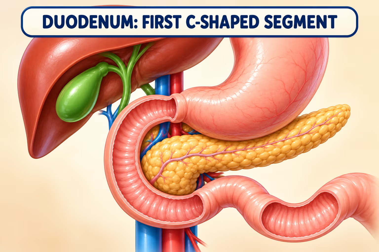
The duodenum is the first, C-shaped segment of the small intestine, continuing from the stomach’s pylorus and curving around the head of the pancreas. It receives chyme from the stomach as well as bile and pancreatic secretions. Bile is produced by the liver and may be stored in the gallbladder; pancreatic ducts carry pancreatic juice. The duodenum mixes these materials and continues digestion. Its hormones also help regulate gastric emptying and acid neutralization.

See NCBI Bookshelf’s duodenum anatomy overview and its pancreas anatomy reference.
What is the dermis?
The dermis is the connective-tissue layer beneath the epidermis. It contains blood and lymphatic vessels, nerves, hair follicles, and glands. Sensory endings in skin detect different stimuli; it is more precise to say that the dermis contains these structures than to describe it as the sole source of every skin sensation or temperature response.
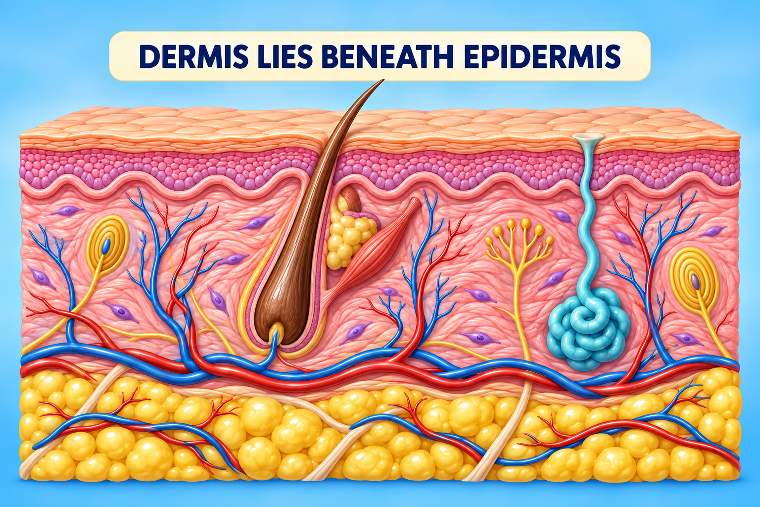
For an overview of skin layers and structures, see the NCBI Bookshelf integument reference.
How do the deltoid muscles move the shoulder?
The deltoid is a large muscle covering the shoulder. Its anterior, middle, and posterior portions contribute differently: the middle portion primarily abducts the arm, while the anterior portion assists flexion and medial rotation and the posterior portion assists extension and lateral rotation. These actions occur with other shoulder muscles, so the deltoid does not work alone.
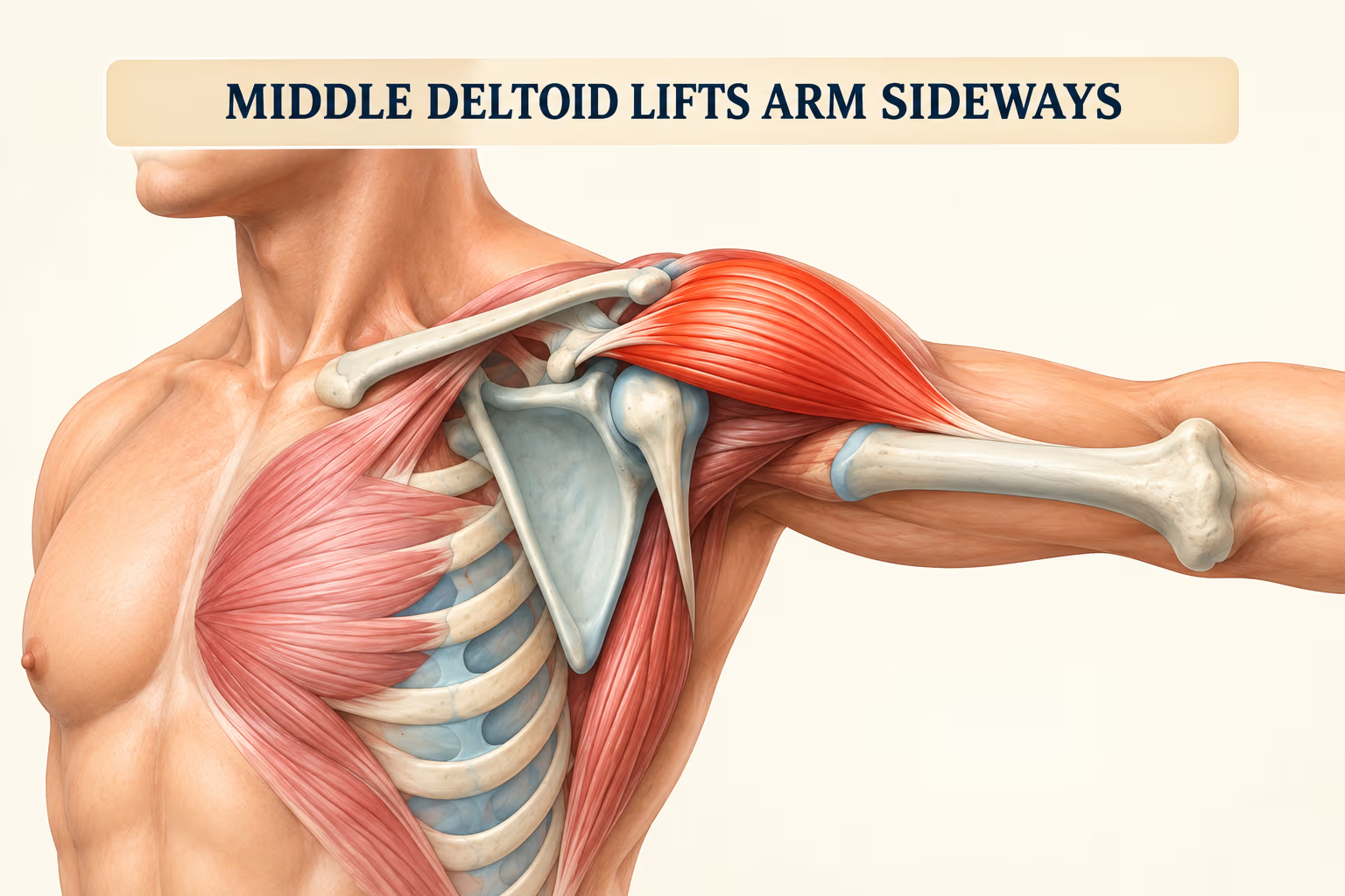
See NCBI Bookshelf’s deltoid muscle anatomy.
What should you know about the diaphragm?
The diaphragm is a dome-shaped muscle and tendon forming the floor of the chest cavity and the roof of the abdominal cavity. When it contracts, it descends and increases the chest cavity’s volume, lowering pressure so air moves into the lungs. It is the primary muscle of quiet inspiration, working with other respiratory muscles.
See NCBI Bookshelf’s diaphragm overview.
How do digital nerves, dendrites, and touch receptors differ?
Digital nerves carry sensory information from the fingers and motor fibers to some hand muscles, depending on the nerve branch. Dendrites are branching extensions of neurons that receive many synaptic inputs; a neuron integrates those inputs, and action potentials typically travel along its axon. Dendrites are microscopic parts of nerve cells, not separate nerves.
Pacinian corpuscles are specialized touch receptors found in deeper skin and other tissues. They are not capsules that surround digital nerves; each corpuscle has layered tissue around a sensory nerve ending and responds especially to vibration and pressure. Touch and temperature are detected by multiple receptor types.
See the NIH brain curriculum overview and NCBI’s sensory-system reference.
What does the ductus arteriosus do before birth?
The ductus arteriosus is a fetal blood vessel connecting the pulmonary trunk to the aorta. It helps route much of the blood away from the fetal lungs, which are not yet carrying out gas exchange. Oxygen enters fetal blood at the placenta and reaches the fetus through the umbilical vein; the ductus arteriosus is a shunt in that circulation, not the route that carries oxygenated blood from the placenta directly to the aorta. After birth, the vessel normally constricts as circulation changes. A persistent ductus arteriosus is a medical condition assessed and managed by clinicians.
See NCBI Bookshelf’s fetal circulation overview and its ductus arteriosus anatomy reference.
How is the dura arranged around the brain and spinal cord?
The dura mater is the tough outermost meningeal membrane. In the skull it has periosteal and meningeal layers; the spinal dura consists of the meningeal layer, with the epidural space outside it. The dural venous sinuses form between the cranial dural layers and drain blood from the brain. The dura supports and surrounds the central nervous system, but it does not consist of three separate layers by itself: dura, arachnoid, and pia are the three meninges.
See NCBI Bookshelf references on cranial meninges and spinal meninges.
What are DNA, ducts, and dental pulp?
DNA is a molecule that stores inherited biological information; it is found in cells and is not an anatomical part like a muscle or organ. Ducts are passageways whose contents and destinations depend on the particular duct. For example, bile ducts carry bile and pancreatic ducts carry pancreatic secretions; bile is made in the liver, although the gallbladder stores and concentrates it.
Dental pulp is the soft connective tissue inside the tooth. It contains blood vessels, nerves, and specialized cells including odontoblasts, which form dentin along the pulp’s boundary. Sensory nerves contribute to pain sensation, but the pulp’s functions include nourishment and dentin support as well.
See NCBI Bookshelf’s biliary duct anatomy and dental pulp reference.
This is a general anatomy learning guide, not a diagnostic or treatment resource.
Original article and D directory
The complete saved source article follows with every original paragraph, heading, table row, list item, link preserved exactly. The explanations above add current anatomic context without rewriting that source.
Dive into our next installment of this captivating anatomical series, “Body Parts That Start With D.” In this article, we explore the fascinating details of body parts starting with the letter ‘D’, such as the dermis or the duodenum.
This informative piece provides insight into the form, function, and unique attributes of these biological components. Ideal for learners, teachers, or anyone curious about the human body, this exploration continues our alphabetical journey through the intricate world of human anatomy. No matter your background, prepare to delve deeper into the complexities of our remarkable bodies.
Human Body Parts That Start With The Letter D
The human body, an incredible marvel of nature, comprises a myriad of parts and structures that play specific roles to maintain life and wellbeing. Continuing our alphabetical exploration of the body, we now reach “D,” a letter that names many fascinating and critical elements of our anatomy. This article will delve into the body parts that begin with “D,” offering insights into their functionality, significance, and their contributions to our overall health.
Duodenum

The duodenum, the first part of the small intestine, plays a vital role in the digestion of food. It is the site where partially digested food from the stomach, called chyme, mixes with bile from the gallbladder and digestive enzymes from the pancreas. This process facilitates the breakdown of food into simpler, absorbable forms. The duodenum also plays a role in regulating stomach acid production to prevent damage to the intestines.
Dermis

The dermis is the layer of skin found beneath the outermost layer, the epidermis. It contains tough connective tissue, hair follicles, and sweat glands. The dermis gives the skin its flexibility and strength, and it is responsible for many of the sensations we feel because it is rich in nerve endings. It also plays an essential role in thermoregulation through sweat production and blood flow regulation.
Deltoids

The deltoids are large, triangular muscles covering the shoulder joints that contribute to the rounded contour of the upper arm. The deltoid muscle is divided into three parts – the anterior, middle, and posterior fibers, each contributing to a different type of arm movement. They are crucial for various movements of the shoulder joint and provide the strength for lifting and rotating the arm.
Diaphragm

The diaphragm is a thin skeletal muscle that sits at the base of the chest and separates the abdomen from the thorax (chest). It plays a critical role in respiration: as it contracts, the volume of the thoracic cavity increases, creating a negative pressure within the chest that draws air into the lungs.
Digits
The term “digits” refers to the fingers and toes in the human body. Each digit, whether on the hand or foot, is an essential tool for touch, grasping, balance, and mobility. The fingers allow for precise manipulation, enabling actions such as writing, typing, and grasping objects, while the toes provide balance and support when standing and walking.
Dendrites

Dendrites are the segments of a neuron that receive stimulation to cause the cell to become active. They conduct electrical messages to the neuron cell body for the cell to function. Dendrites are essential for neural communication, and their structure can be influenced by the activity or lack thereof, playing a role in learning and memory.
Dura Mater

The dura mater is the outermost of the three layers of the meninges that surround the brain and spinal cord. This thick and dense membrane provides a protective coat, helps support the brain, and plays a significant role in supplying blood to the skull and the scalp.
DNA

Although not a “body part” in the traditional sense, DNA (Deoxyribonucleic Acid) is a crucial component of all human cells, and thus warrants a mention. DNA contains the genetic instructions for the development, functioning, growth, and reproduction of all known organisms. It carries the information necessary to build and maintain an organism, in the form of genes.
Ducts

Ducts in the human body are tube-like passages that transport substances from one place to another. Some examples include bile ducts, which carry bile from the liver and gallbladder to the duodenum, and the pancreatic duct, which takes digestive enzymes from the pancreas to the same location. Ducts play a critical role in many bodily functions, particularly in digestion and waste disposal.
Digital Nerves: Feeling the World at Your Fingertips
Imagine tiny filaments woven into your fingers, sending messages of touch, temperature, and pain to your brain – that’s the wonder of the digital nerves. These delicate threads, found in each fingertip, play a crucial role in our experience of the world, allowing us to explore, manipulate, and connect with our surroundings.
Hand showing the location of the major digital nerves in the fingers
- Branching Out: Each digital nerve is a branch of the median nerve or ulnar nerve, reaching the fingertips through the palm and fingers. They further divide into countless tiny branches, ensuring every corner of your fingertip is richly innervated.
- Sensory Symphony: Digital nerves carry various types of sensory information:
- Touch: They allow you to feel delicate textures, pressure, and even the slightest brush against your skin.
- Temperature: Digital nerves sense hot and cold, helping you adjust your grip or avoid uncomfortable temperatures.
- Pain: If you bump your finger, pain signals travel through the digital nerves, alerting your brain to potential harm.
- Protecting the Precious: Tiny capsules called Pacinian corpuscles surround the digital nerves in your fingertips, enhancing their sensitivity to touch and vibration. This delicate system allows you to feel even the subtlest textures and textures, essential for activities like reading Braille or playing a musical instrument.
Ductus: A Bridge for Baby Hearts

Imagine a temporary bypass connecting two chambers of a newborn’s heart – that’s the remarkable ductus arteriosus. This crucial structure plays a vital role in fetal circulation, ensuring optimal oxygen supply to the baby before birth.
Fetal heart showing the location and function of the ductus arteriosus
- Life Before Lungs: Before birth, the baby receives oxygen-rich blood from the placenta through the umbilical cord. The ductus arteriosus bypasses the uninflated lungs, channeling this blood directly to the aorta, supplying vital oxygen to the body.
- Closure Countdown: After birth, the first breaths inflate the baby’s lungs, allowing them to take in oxygen directly. In a healthy baby, the ductus arteriosus naturally constricts and closes within the first few days or weeks of life. This closure creates the adult anatomy with separate pulmonary and systemic circulation.
- Medical Marvels: Sometimes, the ductus remains partially open, requiring medical intervention. However, modern medicine offers safe and effective procedures to close the ductus and ensure proper blood flow in newborns.
Dura: The Brain’s Bodyguard

Imagine a tough, fibrous membrane encasing and protecting your brain – that’s the dura mater. This sturdy shield, the outermost layer of the meninges, plays a crucial role in safeguarding our central nervous system from harm.
Meninges showing the dura mater surrounding the brain and spinal cord
- Multilayered Might: The dura mater is not one, but three layers:
- Dura mater externa: The toughest layer, attached to the skull and providing structural support.
- Dura mater interna: Lines the skull and forms the dural sinuses, carrying blood away from the brain.
- Meningeal layer: Lines the brain and spinal cord, providing a smooth surface and additional protection.
- A Shield and a Support: The dura mater protects the brain from physical injury, infections, and pressure changes. It also supports the brain within the skull, preventing it from sagging or shifting.
- Beyond Brawn: The dura mater plays a vital role in maintaining the cerebrospinal fluid, a protective liquid surrounding the brain and spinal cord. It also contributes to venous drainage and immune function within the central nervous system.
Dental pulp

The dental pulp is the part in the center of a tooth made up of living connective tissue and cells called odontoblasts. The pulp is a critical component of tooth health, containing the blood vessels, nerves, and lymphatic vessels, responsible for nourishing the tooth and sensing pain.
List of Human Body Parts Starting with D
| Descending Aorta | Diaphragm | Digital Nerves |
| Ductus | Dura | Dura, Cranial |
| Dura, Spinal | Duodenum | Dermis |
| Dental pulp | DNA | Dura Mater |
| Dendrites | Deltoids | Digits |
| Ducts |
Conclusion
The journey through the parts of the human body beginning with “D” has revealed several critical components, from the duodenum, vital for digestion, to the dendrites, essential for neural communication. The exploration underscores the complexity of the human body and the myriad pieces that work in harmony to keep us alive and well. It truly is a remarkable reminder of the intricate network of structures and systems that shape our physical existence.
Human Body Parts That Start With
A | B | C | D | E | F | G | H | I | J | K | L | M | N | O | P | Q | R | S | T | U | V | W